Research
Our lab’s goal is to phenotype mitochondria in both healthy and diseased states. We specialize in basic molecular biology techniques, skeletal fiber bundle separation, respirometry (Oroboros O2K), mitochondrial membrane potential, H2O2 emission, indices of cellular redox state, and other methods to measure mitochondrial bioenergetic parameters. We study cellular, rodent, and human models of disease.
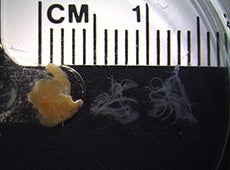
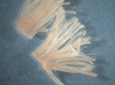
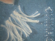
Skeletal fiber bundle separation


H2O2 emission via Amplex Ultra Red in a Horiba Fluorometer
Permeabilized fibre bundles were prepared from red portions of the gastrocnemius muscle from C57BL/6N (6N) or C57BL/6J (6J) mice. Representative trace of H2O2 emission during respiration supported by sequential addition of pyruvate (Pyr) and carnitine (Carn) in fibres from 6N (+NNT) compared with 6J (−NNT) mice. Biochem. J. (2015) 467, 271–280 (Printed in Great Britain) doi:10.1042/BJ20141447
Mitochondrial membrane potential via an ion selected electrode with corresponding respiration in the Oroboros O2K
Mitochondria were isolated from gastrocnemius and quadriceps muscle from C57BL/6N or C57BL/6J mice. Direct comparison of proton conductance in isolated mitochondria from C57BL/6N and C57BL/6J mice under substrate conditions inducing maximal H2O2 production from PDHC and complex I (succinate + pyruvate + carnitine + glutamate). Note greater proton conductance in C57BL/6N mice at the highest common ΔΨm. Car, carnitine; Glu, glutamate; Pyr, pyruvate; Suc, succinate. Biochem. J. (2015) 467, 271–280 (Printed in Great Britain) doi:10.1042/BJ20141447
Proton Leak Assay trace
TPP titration was done first to create a standard curve of [TPP]. Isolated mitochondria were introduced with the presence of energetic substrates. The decreasing of the [TPP] measured in the buffer indicates the uptake of TPP by mitochondria due to high mitochondria membrane potential. Malonate titration was followed to inhibit complex II, thus the reduction of membrane potential and respiration rate. Back trace: [TPP]; Blue trace: [O2]; Red trace: JO2
